

Advanced Magnetic Resonance Elastography for the Brain
Project period
January 2024 – December 2025
Objective
This project aims to combine magnetic resonance elastography (MRE) and advanced diffusion magnetic resonance imaging (dMRI) to develop the next generation of MRE methods to improve the biomechanical characterization of brain tissue.
More specifically, A) we will propose computational models that consider the diffusion processes to estimate biomechanical properties of the brain at a sub-voxel level and B) we will use advanced dMRI data to predict biomechanical properties without the expensive equipment required by MRE.
We will apply these new methods to data we are acquiring from Parkinson’s disease (PD) and cancer patients and healthy subjects and test their potential to improve the diagnosis and treatment of patients and to characterize biomechanical changes during ageing.
Background
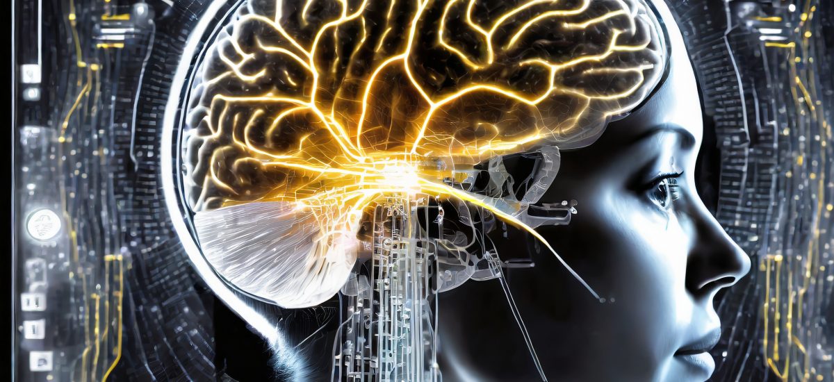
Neurological disorders affect millions of persons worldwide. While MRI is crucial in clinical diagnostics, it is largely limited to morphological features. Understanding how the mechanical properties of tissues change due to disease can give valuable information for improving their early detection and diagnosis. A promising tool for non-invasively estimating these mechanical properties through magnetic resonance imaging (MRI) is Magnetic Resonance Elastography (MRE). The current MRE methods generate limited mechanical information about the brain tissue at a relatively low image resolution. The proposed methods will provide a better mechanical characterization of tissues for improved diagnosis. Moreover, using additional MRI modalities, we can better understand the mechanism behind the changes in mechanical properties due to diseases. This can eventually lead to better treatments for patients. MRE for the brain is only available in a few sites worldwide. In Autumn 2022, the KTH-owned MRI scanner became the first one in Sweden capable of performing MRE examinations in the brain.
Crossdisciplinary collaboration
In this project, we are establishing a new cross-disciplinary and complementary collaboration:
- Lisa Prahl Wittberg, Prof. in Multiphase flows / Fluid mechanics at the Department of Engineering Mechanics at KTH, is an expert in developing computational models to understand better the underlying processes of complex fluids in the human body.
- Rodrigo Moreno, Assoc. Prof. in Biomedical Imaging at KTH is an expert in using advanced AI applied to medical images. His group is currently involved in all projects in Sweden that use MRE for the brain.
- Christian Gasser, Prof. in tissue mechanics at KTH Mechanical Engineering, provides his expertise in the constitutive modelling of tissues needed in the project.
- Christoffer Olsson, a postdoc recruited for the previous phase of the project, will remain one of the key persons for developing the methods in this project.
The data of this project is being collected in collaboration with Assoc. Prof. Armita Golkar from Stockholm Univ (SU), and Prof. Per Svenningsson, Assoc. Prof. Grégoria Kalpouzos and Assoc. Prof. Anna Falck Delgado from Karolinska Institute (KI).
Contacts

Rodrigo Moreno
Associate Professor, School of Engineering Sciences in Chemistry, Biotechnology and Health (CBH) at KTH, Co-PI of project Characterization of the mechanical tissue properties of the brain in the developing brain with magnetic resonance elastography, Co-PI of project Advanced Magnetic Resonance Elastography for the Brain, Digital Futures Faculty
+46 8 790 9787rodmore@kth.se

Lisa Prahl Wittberg
Professor, Department of Engineering Mechanics at KTH, Co-PI of project Advanced Magnetic Resonance Elastography for the Brain
+46 8 790 75 80prahl@mech.kth.se