

About the project
Objective
Cancer progression is driven not only by genetic alterations within individual cells but also by complex communication between cancer cells and their surrounding microenvironment. Understanding and controlling these interactions would enable therapies that disrupt cancer-promoting cellular equilibria, improve treatment design, and accelerate precision medicine. The major technical challenge is to develop predictive computational models that capture high-dimensional, nonlinear cell-cell interactions and reliably forecast the effects of targeted interventions. A promising path to address this challenge is to combine biological knowledge on cellular signaling and regulatory mechanisms with recent advances in AI and dynamical systems theory. This would entail learning biologically consistent models of multicellular behavior, validated and enriched on micro-physiological systems.
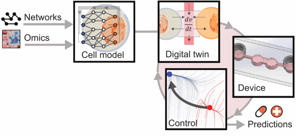
As a first approach, we focus on communication between pancreatic cancer cells and stromal cells, whose reciprocal interactions are known to reinforce tumor growth and therapeutic resistance. While recent single-cell technologies provide unprecedented measurements of cellular states and signaling pathways, these data remain largely observational and therefore have limited predictive power for designing interventions. Moreover, existing AI approaches rarely account for the coupled dynamics governing cellular communication, making the identification of stable and controllable cellular states particularly challenging. This motivates our project, SCALE, to develop biology-informed AI methods for modeling and controlling multicellular systems. The objectives of SCALE are to establish the fundamental theory and computational tools needed to learn predictive models of cell-cell interactions, to identify healthy and disease-associated equilibria, and to design optimal in silico interventions to guide experimental validation and future therapeutic development.
Background
Cells continuously exchange signaling molecules to create the cellular microenvironment. In pancreatic cancer, communication between cancer cells and surrounding stromal cells regulates processes such as tumor growth and therapeutic resistance. In reality, these cell-cell interactions are not directly observed. Instead, they are studied through molecular measurements and cell culture experiments that capture the cellular states resulting from these interactions. Therefore, it is difficult to directly characterize the interaction mechanisms and predict how perturbations of one cell population influence the others.
The functional relationship between cell-cell interactions and the observed cellular states is governed by complex biological processes involving intracellular signaling and intercellular communication. For multicellular systems such as the pancreatic tumor microenvironment, these coupled dynamics remain difficult to characterize because of the large number of interacting molecules and feedback mechanisms. Furthermore, experimental observations are inherently noisy and capture only part of the underlying biological processes.
Naturally, modeling and predicting the coupled dynamics that govern cell-cell interactions from experimental observations constitute a challenging systems modeling problem.
Cross-disciplinary collaboration
The project will combine methods and techniques from separate research fields – (a) biological knowledge about virtual cell models and computational biology, (b) dynamical system and control theories, (c) micro physiological systems design, and experiments.
About the project
Objective
Our project will use AI to effectively turn current methods for two-dimensional (2D) spatially resolved molecular methodologies into three-dimensional (3D) methodologies and allow molecular studies of tissues as 3D objects. The Spatial Transcriptomics methodology, commercially available as Visium, is the most widely used technique to measure gene expression in 2D tissue sections, and with the recent release of VisiumHD, gene expression analysis can be obtained at unprecedented subcellular spatial resolution. We will create AI algorithms necessary to synthesize, analyze, and visualize 3D representations of cancer tissue that otherwise would be experimentally prohibitively expensive. We will exploit deep learning and construct 3D models using advanced AI methodologies such as Diffusion Models, Flow Matching, Schrödinger Bridges, and Optimal Transport methods. Specifically, we will infer 3D distributions of cell quantities from 2D data and generate continuous 3D reconstructions of tumor and metastatic tissue.
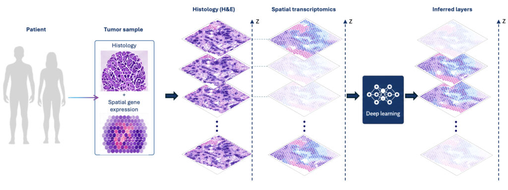
Our vision is to develop general AI methods required for revealing and comparing the 3D spatial organization of tumor tissues at reduced cost. The methods will be showcased by application to patient-matched primary tumor and metastasis samples from breast and prostate cancer patients, providing important understanding of the metastasis process, seeding patterns, and differences between primary and metastatic tumor micro environments.
Background
Around 90% of all cancer patients die due to metastases, colonies of cancer cells that have established themselves at a distance from the primary tumor. Despite this, metastases are surprisingly understudied and consequently not well-understood. Most patients with breast or prostate cancer are diagnosed with localized disease. However, when the disease has spread and created metastases, effective treatments typically do not exist. This highlights the necessity of new, more efficient therapies aimed toward clinically established metastases as well as micro-metastases with growth potential.
Spatially resolved gene expression data have transformed biomedical research and motivated extensive method development for mapping cell types and integrating multiple slides and molecular modalities. However, these approaches remain restricted to 2D views of tissue, and understanding the 3D organization of tumors and their microenvironments represents a major challenge. Current spatial omics and lineage analyses remain 2D. By combining cutting-edge biological data with state-of-the-art AI methodologies for 3D reconstruction and lineage inference, this project advances computational methods and biological understanding of tumor evolution and metastatic dissemination.
Cross-disciplinary collaboration
The project integrates AI and computational modeling with spatial omics in the context of clinical cancer biology. The groups combine expertise in machine learning and probabilistic modeling for inference and synthesis of spatial and lineage data, together with experimental spatial omics and single-cell sequencing. Clinical collaborators will guide the biological questions, ensuring relevance to metastatic disease and tumor microenvironment differences. The work is organized into three components: data generation, computational methods development (2D analysis and 3D synthesis), and biological investigation.
The computational framework integrates single-cell, spatial, and imaging data to enable 3D spatial modeling of tumor evolution. Clinically important questions, centered on metastatic potential, clonal seeding patterns, and tumor–microenvironment interactions, will drive the biological investigation. Through joint supervision, shared infrastructure at SciLifeLab and collaboration with national and international stakeholders, the project establishes a translational bridge from computational methods to clinical application and contributes to precision oncology.
About the project
Objective
The goal of this project is to better understand how the brain changes in diseases like Alzheimer and Parkinson. We study the brain’s wiring using a special type of MRI scan and look for patterns in how different regions are connected. By using advanced mathematical tools, we can detect subtle changes in these brain networks that are often missed by standard methods. These patterns may help us distinguish between healthy and diseased brains, and even identify early signs of disease before symptoms become clear. Ultimately, this research could lead to more accurate and earlier diagnosis, as well as a better understanding of how these diseases develop over time.
Background
Alzheimer’s and Parkinson’s diseases are the two most common neurodegenerative diseases, affecting more than 50 million people worldwide. They have a major impact not only on the patients, but also their families and healthcare systems. By improving early diagnosis through advancing methods to learn from complex brain connectivity patterns, this proposal has the potential to generate, in the future, benefits that extend beyond the scientific community, contributing both to societal well-being and to the sustainability of healthcare systems.
Tractography from diffusion magnetic resonance imaging (dMRI) reveals the wiring of the human brain but remains challenging to analyze due to its complexity and variability. Classic approaches, such as streamline count, fractional anisotropy, mean diffusivity, and tract length often capture pairwise relationships, but neglect the higher-order structural organization of the white matter network. As a result, complex and spatially distributed alterations that emerge early in neurodegenerative diseases can remain undetected.
Topological data analysis (TDA) and machine learning on graphs offer promising complementary tools to address this limitation. TDA provides tools to quantify the global and local topology of structural connectivity, such as loops, cycles, and motifs, that go beyond metrics accounting for pairwise interactions. Graph-based machine learning methods, such as graph neuronal network (GNN), can then leverage this richer representation to learn complex patterns of connectivity and distinguish between healthy and diseased brains.
About the Digital Futures Postdoc Fellow
Ilaria Carannante is a Computational Neuroscientist with a background in mathematics. She holds a PhD in Computer Science, with specialization in Computational Biology from KTH. Ilaria has always been passionate about science and mathematics, and her biggest career goal is to contribute expertise in neuroscience to advance our understanding of the human brain and to develop approaches for remedying disorders that impede its proper functioning.
During her PhD in Professor Jeanette Hellgren Kotaleski’s lab, she developed data-driven multiscale models of a brain region called Striatum. As a postdoctoral researcher in Professor Alain Destexhe’s group at CNRS in Paris, she expanded her work to larger-scale networks with a special focus on the Basal Ganglia (to which the striatum belongs). The next natural step in her research journey is to extend this multiscale approach to the human brain. For this reason in her current research she aims to leverage diffusion MRI, combined with topological data analysis and graph neural networks, to uncover higher-order patterns in brain connectivity.
In the long term, she aims to build data-driven whole-brain models to better understand brain function in health and Parkinson’s disease, with the ultimate goal of improving diagnosis and guiding targeted interventions.
Main supervisor
Martina Scolamiero, Associate Professor at the division of Algebra, Combinatorics, Geometry and Topology, KTH.
Co-supervisor
Rodrigo Moreno, Professor at the division of Biomedical Imaging, KTH.
About the project
Objective
This project aims to implement intrinsic magnetic resonance elastography (I-MRE). Instead of the pump, it uses vascular pulsatility as the source of tissue movement. More specifically, A) we will adapt and test I-MRE techniques to acquire subtle brain tissue motion in MRI scanners; B) develop and validate machine learning and computational models to estimate mechanics from I-MRE data; C) inform traumatic brain injury (TBI) simulations with I-MRE.
Background
Magnetic resonance elastography (MRE) in the brain is a new technique in which the mechanical properties of the brain tissue can be estimated non-invasively. Unfortunately, the standard MRE setting requires an expensive pneumatic pump and specialized software, hindering its use in most hospitals.

Cross-disciplinary collaboration
This project is creating a new multidisciplinary collaboration between three PIs with complementary expertise in understanding the biomechanical properties of the brain: Rodrigo Moreno’s group is an expert in magnetic resonance elastography. Zhou Zhou’s group’s expertise is in neuroimaging-informed finite-element simulation of brain biomechanics. Lisa Prahl Wittberg’s research is focused on computational fluid mechanics (CFD)blood flow dynamics using both experimental and numerical methods (computational fluid mechanics, CFD).
About the project
Objective
- Careful assessment of what new information due to personal data of an individual has been memorised;
- Guidelines outlining what residuals of personal information in a ML model should be legally acceptable;
- Explore how the RTBF can be effectively implemented, ultimately in the context of ML systems.
Background
In the context of machine learning (ML), there is an urgent need to clarify what “deletion” under the Right to be Forgotten (RTBF) truly entails. As ML models generalize and internalize patterns from data, achieving complete removal of an individual data point remains a major technical challenge. We argue, however, that such full deletion often exceeds what the law actually requires. REFLECT-ML aims to explore how the RTBF can be effectively implemented, ultimately in the context of ML systems. We will address the disconnect between the abstract legal language of regulators and operationally founded technical theoretical measures that can be used to quantify the new information associated with individual data as well as the practical complexities in estimating those. Further, we will develop different technical approaches to control its memorization as well as exploring machine unlearning attempts.
Cross-disciplinary collaboration
The team consists of PI Oechtering (researcher in information theory, relevant for information quantification), PI Colonna (researcher in law, relevant knowledge related to the GDPR and AI Act), and PI Johansson (researcher in optimization, relevant for the design of efficient learning algorithms). The outlined work is a cross-disciplinary effort since information measures need to be legal compliant, legal requests need to be algorithmically feasible, and algorithms need to aim for the right objective.
About the project
Objective
This project aims to establish an AI-based online platform for automated, and robust personalization and positioning of HBMs, focusing on infant HBMs. By developing a family of infant HBMs equipped with efficient personalization and a novel AI-based positioning pipeline, the project facilities rapid and subject-specific model generation that can foster industrial and clinical innovations relating to infant safety.
Background
Finite element human body models (HBMs) are digitalized representations of the human body and have become essential tools in both industrial innovation and clinical applications. These models often are a baseline and in a specified position, and before the use of the HBMs, personalization and positioning of HBMs are needed. Despite continuous active development, HBM positioning remains challenging and tedious; further comparing with existing adult HBMs, infant and child HBMs are underdeveloped.
This project builds on, and further develops, the results from the Research Pair project: “AI-based Positioning and Personalization Platform for Human Body Models (HBMs)“.
Crossdisciplinary collaboration
This project combines expertise within mechanical and biomechanical modeling (from KTH School of Engineering Sciences in Chemistry, Biotechnology and Health) with expertise in artificial intelligence (from the Department of Industrial Systems at RISE).
About the project
Objective
The objective of this project is to develop an AI-enabled, fully self-powered, and biodegradable wound-healing patch that accelerates tissue regeneration and enables continuous monitoring of post-cardiac-surgery wounds. The system combines triboelectric nanogenerators (TENGs) for bioelectric stimulation with AI-based image analysis to provide personalized, sustainable, and real-time wound care without external power sources.
Background
Post-cardiac-surgery wounds face high risks of infection, delayed healing, and limited continuous monitoring. Existing wound-care technologies rely on external power sources, are costly, and lack portability. Triboelectric nanogenerators (TENGs) offer a promising alternative by harvesting biomechanical energy from natural body movements to deliver gentle bioelectric stimulation.
This project integrates biodegradable hydrogels with antibacterial and anti-inflammatory properties and AI-driven wound image analysis to assess healing stages such as inflammation, proliferation, and remodeling. The approach reduces electronic waste, enables continuous monitoring, and supports faster, safer recovery through sustainable digital healthcare solutions.
About the Digital Futures Postdoc Fellow
Swati Panda is a postdoctoral researcher at the Department of Biomedical Engineering and Health Systems at KTH, specializing in biocompatible and biodegradable energy-harvesting devices for self-powered healthcare applications. She holds a PhD in Robotics and Mechatronics Engineering from DGIST, South Korea. Her research focuses on triboelectric and piezoelectric nanogenerators, biodegradable/biocompatible polymers, and AI-based signal and image processing for healthcare sensing.
She has extensive experience in material synthesis, device fabrication, in-vivo experimentation, and self-powered health monitoring systems. Through her work, she aims to develop sustainable, wearable, and smart healthcare technologies that improve patient outcomes while reducing environmental impact.
Main supervisor
Seraina Dual, Assistant Professor, Department of Biomedical Engineering and Health Systems, KTH Royal Institute of Technology.
Co-supervisor
Erica Zeglio, Assistant Professor, Department of Chemistry, Stockholm University.