About the project
Objective
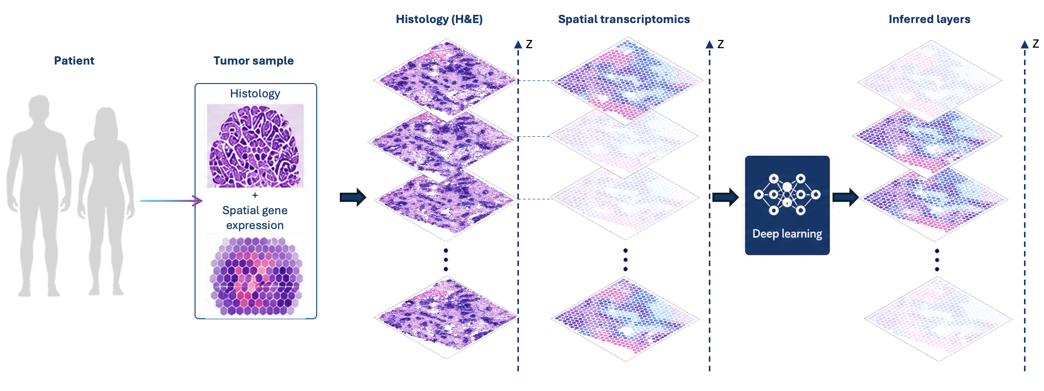
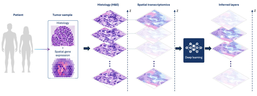
Our project will use AI to effectively turn current methods for two-dimensional (2D) spatially resolved molecular methodologies into three-dimensional (3D) methodologies and allow molecular studies of tissues as 3D objects. The Spatial Transcriptomics methodology, commercially available as Visium, is the most widely used technique to measure gene expression in 2D tissue sections, and with the recent release of VisiumHD, gene expression analysis can be obtained at unprecedented subcellular spatial resolution. We will create AI algorithms necessary to synthesize, analyze, and visualize 3D representations of cancer tissue that otherwise would be experimentally prohibitively expensive. We will exploit deep learning and construct 3D models using advanced AI methodologies such as Diffusion Models, Flow Matching, Schrödinger Bridges, and Optimal Transport methods. Specifically, we will infer 3D distributions of cell quantities from 2D data and generate continuous 3D reconstructions of tumor and metastatic tissue.
Our vision is to develop general AI methods required for revealing and comparing the 3D spatial organization of tumor tissues at reduced cost. The methods will be showcased by application to patient-matched primary tumor and metastasis samples from breast and prostate cancer patients, providing important understanding of the metastasis process, seeding patterns, and differences between primary and metastatic tumor micro environments.
Background
Around 90% of all cancer patients die due to metastases, colonies of cancer cells that have established themselves at a distance from the primary tumor. Despite this, metastases are surprisingly understudied and consequently not well-understood. Most patients with breast or prostate cancer are diagnosed with localized disease. However, when the disease has spread and created metastases, effective treatments typically do not exist. This highlights the necessity of new, more efficient therapies aimed toward clinically established metastases as well as micro-metastases with growth potential.
Spatially resolved gene expression data have transformed biomedical research and motivated extensive method development for mapping cell types and integrating multiple slides and molecular modalities. However, these approaches remain restricted to 2D views of tissue, and understanding the 3D organization of tumors and their microenvironments represents a major challenge. Current spatial omics and lineage analyses remain 2D. By combining cutting-edge biological data with state-of-the-art AI methodologies for 3D reconstruction and lineage inference, this project advances computational methods and biological understanding of tumor evolution and metastatic dissemination.
Cross-disciplinary collaboration
The project integrates AI and computational modeling with spatial omics in the context of clinical cancer biology. The groups combine expertise in machine learning and probabilistic modeling for inference and synthesis of spatial and lineage data, together with experimental spatial omics and single-cell sequencing. Clinical collaborators will guide the biological questions, ensuring relevance to metastatic disease and tumor microenvironment differences. The work is organized into three components: data generation, computational methods development (2D analysis and 3D synthesis), and biological investigation.
The computational framework integrates single-cell, spatial, and imaging data to enable 3D spatial modeling of tumor evolution. Clinically important questions, centered on metastatic potential, clonal seeding patterns, and tumor–microenvironment interactions, will drive the biological investigation. Through joint supervision, shared infrastructure at SciLifeLab and collaboration with national and international stakeholders, the project establishes a translational bridge from computational methods to clinical application and contributes to precision oncology.