

Spatiotemporal reconstruction with learned deformations for earlier cancer detection via PET imaging
Objective
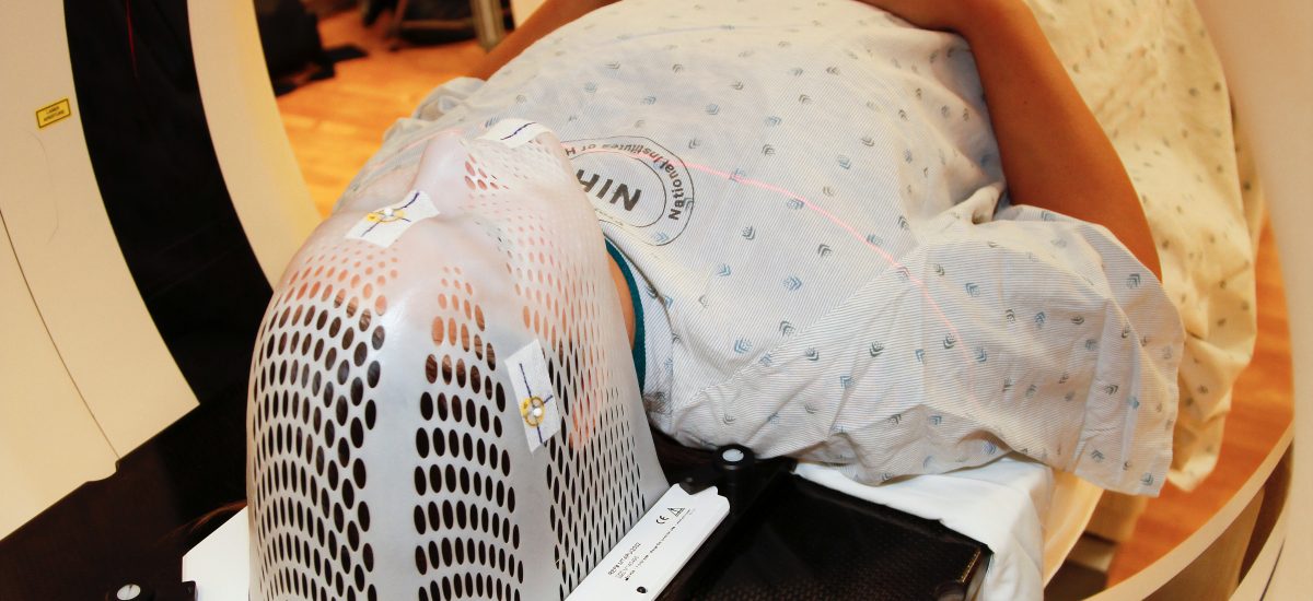
The main aim of this project is to increase the diagnostic power of PET images for detecting lung cancer lesions at an early stage by overcoming the loss of contrast and spatial resolution caused by respiratory motion during data acquisition. Traditional ways of tackling this problem are computationally too demanding to be useful in clinical practice. For this reason, we will implement algorithms deploying a mixture of modelling of the data-acquisition set-up and machine-learning-based tools for image registration. The challenges of this project consist mainly in tackling the size of this four-dimensional image reconstruction problem and in being able to deliver images to radiologists in a time frame compatible with the hospital workflow.
In collaboration with our clinical partners at Karolinska Hospital in Huddinge, we will collect gated PET line-of-response activity data and corresponding 3D CT attenuation maps of the thorax region of 400 patients and test and optimise our 4D algorithms and gating strategies on this data. We will evaluate the resulting reconstructions with a particular focus on their diagnostic power for small lung lesions and make the reconstruction software package openly available.
Background
PET (Positron Emission Tomography) is a medical imaging modality that reconstructs the 3D distribution of metabolic activity by detecting photons emitted during the in vivo annihilation of free electrons with positrons from an injected radioactive tracer. In principle, cancer lesions will be visible in the reconstructed image with high contrast compared to the surrounding healthy tissue thanks to their peculiar metabolic fingerprints (e.g. higher sugar metabolism). PET is, in fact, one of the most powerful imaging modalities for cancer diagnosis and staging. However, the long acquisition time required to collect projection data with an acceptable noise level leads to motion artefacts that strongly affect the contrast-to-noise-ratio of lesions. This is of particular interest when trying to detect tumours that are of a size comparable to the system resolution (~5 mm) and that are continuously moving because of respiratory motion. Those lesions are the most important ones to detect for early-stage diagnosis, leading to a better prognosis for the patient.
Crossdisciplinary collaboration
This is a project in which state-of-the-art mathematical research for solving large inverse problems meets the clinical practice of medical imaging and brings together faculty from the Department of Mathematics at the SCI school at KTH with faculty from the Department of Biomedical Imaging of the CBH school.
Watch the recorded presentation at the Digitalize in Stockholm 2023 event:
Contacts
Massimiliano Colarieti Tosti
Associate Professor, Division of Biomedical Engineering at KTH, Working group Educational Transformation, PI of project Spatiotemporal reconstruction with learned deformations for earlier cancer detection via PET imaging, Digital Futures Faculty
+46 8 790 48 61mct@kth.se
Ozan Öktem
Associate Professor at KTH, Co-PI of project Spatiotemporal reconstruction with learned deformations for earlier cancer detection via PET imaging
ozan@kth.se